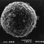
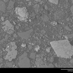
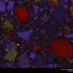
Scanning Electron Microscopy (SEM) uses electrons to raster a sample’s surface showing topography, Z-contrast (phase composition differentials), and chemistry at high magnification.
Energy Dispersive Spectroscopy (EDS) chemistry can be utilized in conjunction with SEM to probe specific phases or create phase maps of area of interest in field of view.
Back-scattered Electron Microscopy Imaging (BSI) shows chemical composition combinations differentials, with bright BSI reflecting high Z (total atomic number composition for a specific phase) compared to dull BSI reflecting low Z.
Raw (unpolished) samples, polished thin sections (1″ x 1-1/2″ x 30 um), and polished sections (1″ diameter) are typical sample inputs for SEM, EDS, and BSI for exploration, economic geology, applied mineralogy, process mineralogy, applied microscopy, and mineral analysis.


